

Abstract
Due to the biocompatibility and biodegradability, as well as the mechanical properties of the fibres, spider silk has become an attractive material for researchers in terms of biomedical applications. In this study, the engineered recombinant spider silk protein eADF4 (C16) was modified with the RGD integrin recognition sequence by a genetic (fusing the GRGDSPG amino acid sequence) and chemical (using cyclic peptide c (RGDfK)) approach. . Both modified silk proteins were processed into films and subsequently characterized for secondary structure, water contact angle, and surface roughness.
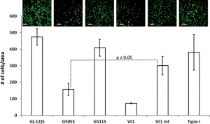
No influence of RGD modifications on any of these film properties could be detected. However, BALB/3T3 mouse fibroblast attachment and proliferation were significantly enhanced in films made from RGD-modified silk proteins. Interestingly, the engineered hybrid protein (with a linear RGD sequence) showed similar or slightly better cell adhesion properties than the chemically modified silk protein with the cyclic RGD peptide.
Description
CUSABIO synthesized the recombinant gene by integrating the N-terminal 6xHis-SUMO tag sequence into the target gene encoding 24-265aa of human EPCAM. The synthesized gene was subsequently cloned into an expression vector. After cloning, the expression vector was introduced into E. coli for expression. The product was purified to obtain recombinant human EPCAM protein bearing the N-terminal 6xHis-SUMO tag. SDS-PAGE analyzed the purity of this recombinant EPCAM protein greater than 90%. This EPCAM protein migrated along with the gel to a band of approximately 43 kDa molecular weight.
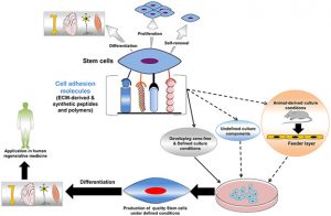
EPCAM is a gene that provides instructions for making a protein called epithelial Cell adhesion Recombinants molecule (also abbreviated as Ep-CAM and CD326) in humans and belongs to the EPCAM family. CD326, a transmembrane glycoprotein, plays an important role in the appearance and development of colorectal cancer. CD326 is a multifunctional molecule, including cell cycle acceleration, promotion of cell proliferation, differentiation, migration, and immune escape. Furthermore, most tumours have EpCAM expression, with most epithelial tumours showing high expression, non-epithelial tumours expressing weakly or not at all, and some mesenchymal tumours only showing weakly positive.
Purity: greater than 90% as determined by SDS-PAGE.
Destination Names: EPCAM
Uniprot No.: P16422
Research area: labels and cell markers
Alternative Names
17 1A; 323/A3; adenocarcinoma-associated antigen; adenocarcinoma-associated antigen; Antigen identified by monoclonal AUA1; AUA1; CD326; CD326 antigen; Trop 1 cell surface glycoprotein; cell surface glycoprotein Trop 2; Trop-1 cell surface glycoprotein; CO17A; CO17 1A; CO17A; DIAR5; PGE 2; EGP; EGP2; EGP314; EGP40; Ep CAM; Ep-CAM; EPCAM; EPCAM_HUMAN; EpCAM1; epithelial cell adhesion molecule; epithelial cell adhesion molecule intracellular domain (EpCAM-ICD); epithelial cell surface antigen; epithelial cell adhesion molecule; epithelial glycoprotein 1; epithelial glycoprotein 314; epithelial glycoprotein; THAT; GA733 1; GA733 2; GA733-2; gastrointestinal tumor-associated antigen 2; 35-KD glycoprotein; gp4; hEGP2; hEGP314; HNPCC8;
human epithelial glycoprotein 2; KS 1/4 antigen; KS1/4; KSA; Ly74; lymphocyte antigen 74; M1S1; M1S2; M4S1; major gastrointestinal tumor-associated protein GA733 2; major gastrointestinal tumor-associated protein GA733-2; mEGP314; Membrane component chromosome 4 surface marker (35 kD glycoprotein); membrane component; chromosome 4; surface marker 1; membrane component; chromosome 4; surface marker; MIC18; MK1; 289A protein; TACD1; TACSTD1; TROP1; tumor-associated calcium signal transducer 1; Precursor of tumor-associated calcium signal transducer 2; Tumor-associated calcium signal transducer 1
Species: Homo sapiens (Human)
Source: E.coli
Expression region: 24-265aa
Mole Weight: 43.4 kDa
Protein length: Partial
Tag information: N-terminal 6xHis-SUMO-tagged
Form: Liquid or Lyophilized Powder
Note: We will preferably ship the format we have in stock, however, if you have any special requirements for the format, please remark your requirement when placing the order, we will prepare according to your demand.
Buffer
If the dosage form is liquid, the default storage buffer is Tris/PBS based buffer, 5%-50% glycerol.
Note: If you have any special requirements for glycerol content, please remark it when you order. If the administration form is a lyophilized powder, the buffer before lyophilization is a Tris/PBS-based buffer, 6% trehalose, pH 8.0.
Reconstitution
We recommend that this vial be briefly centrifuged before opening to bring the contents to the bottom. Reconstitute protein in sterile deionized water at a concentration of 0.1-1.0 mg/mL. We recommend adding 5-50% glycerol (final concentration) and an aliquot for long-term storage at -20°C/-80°C. Our final default glycerol concentration is 50%. Customers could use it for reference.
Storage conditions
Store at -20°C/-80°C upon receipt; it is necessary to divide it into aliquots for multiple uses. Avoid repeated cycles of freezing and thawing.
Shelf life
Shelf life is related to many factors, storage condition, buffer ingredients, storage temperature and the stability of the protein itself. Generally, the shelf life of the liquid form is 6 months at -20°C/-80°C. The shelf life of the lyophilized form is 12 months at -20°C/-80°C.
Delivery time: 3-7 business days
Notes: Repeated freezing and thawing is not recommended. Store working aliquots at 4°C for up to one week.